Doctors say that this AI tool can find hidden breast cancers
The new tool can be vital for women with dense breast tissues.
The National Breast Cancer Foundation believes that One woman in eight In the United States Risk of four to six times cancer. With the progress of technology and research, we know much more about mammography relationships and risk factors than ever before. And now, a new AI tool could help locate breast cancer in women with dense fabrics that standard images do not always detect.
In relation: Scientists may have cracked the code to predict breast cancer in dense tissues .
A new AI tool can more precisely locate the tumors of the cancerous breast.
In a study published in the journal Radiology , the researchers have unveiled a new AI detection model which can precisely locate cancer tumors on breast images. The tool does not replace magnetic resonance imaging (MRI), but could be used to "improve the accuracy and efficiency of mammary MRI screening", according to the authors.
"AI assisted MRI could potentially detect cancers that humans would not find otherwise", " Felipe Oviedo , PHD, main study author and main research analyst at Microsoft’s IA for Good Lab, said in A press release .
But if the MRIs are more complete than mammograms, they are more expensive and have a higher false rate. Researchers think that the new AI detection model could fill the gap and promote the progression of women's health.
Most importantly, perhaps, the AI model could be a backup tool for women with dense breast tissue, a factor that increases the risk of breast cancer, according to the Breast Cancer Research Foundation (BCRF).
In relation: Doctors say that most women over 65 do not need PAP smear - cancer prices say the opposite .
The test could be vital for women with dense breast tissues.
As Best life Previously, "dense mammary fabric refers to breasts composed of less fatty fabric and more fibrous and glandular fabric - in other words, this is how your breasts appear on a mammography. You cannot feel dense breast tissue, and that does not cause any pain or change in the breast. ”
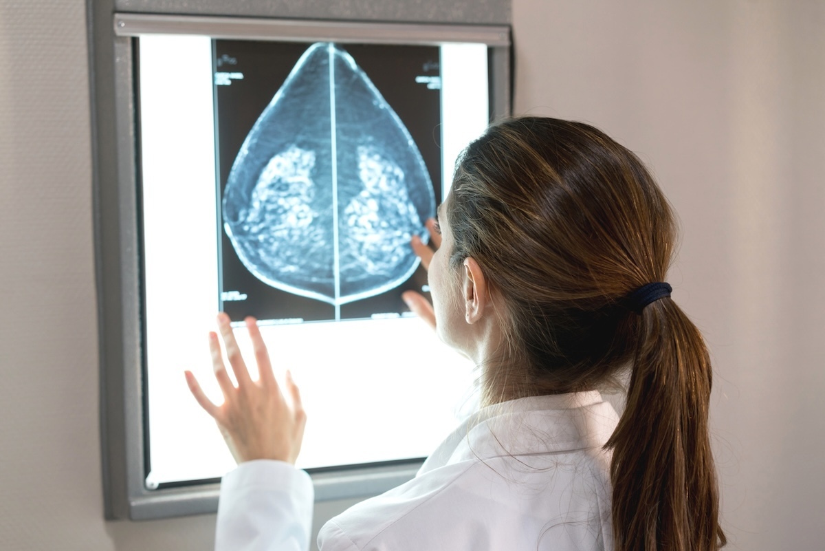
On a mammography, dense breast tissues and tumors appear white, which (it goes without saying) makes it more difficult for radiologists to decipher what is potentially cancerous. It can also be more difficult to see tumors through dense fabrics, adds Cleveland clinic .
Breast cancer begins in your fibroglandular fabric, which is naturally dense. Unfortunately, "the more fibroglander you have in your breast, the more breast cancer you will develop the clinic.
There are four types of dense breast tissue:
- Mainly fatty tissue
- Dispersed fibroglendular breast tissue
- Heterogeneous dense breast fabric
- Extremely dense breast fabric
Of the four, the dispersed fibroglandular and heterogeneous breast tissues are the most common (40% of women).
"Dense breast tissues are more frequent in women under 40 Best life .
In relation: 85% of non -vaccinated women will probably get this virus - and a new research connects it to heart disease .
How does the tool work and when is it available?
The AI anomaly detection model is an original idea of the Olviedo research team and clinical investigators in the Radiology Department of Washington University. It is designed to distinguish normal and abnormal data and reporting anomalies for more in -depth assessment. Consequently, the MRI images receive an "estimated anomaly score".
So how did they achieve this? The researchers formed the AI model "using data of nearly 10,000 consecutive contrast exams", including 42.9% with heterogeneous dense breasts and 11.6% with extremely dense breasts.
Oviedo called the AI tool "a promising solution".
"Unlike traditional binary classification models, our anomalies detection model has learned a solid representation of mild cases to better identify abnormal malignant tumors," he said.
In addition, researchers said that the AI tool can produce "a spatially resolved thermal card" for MRIs. With this functionality, the patterns of the thermal card or the "colors" regions of the image of the breast it detects are abnormal.
This characteristic was tested on 171 women who received MRI for screening or preoperative evaluation for known cancer and 221 women with invasive breast cancer. According to the results, "the abnormal regions identified by the paired model of the malignant tums proven of biopsy annotated by a radiologist, far beyond the performance of the reference models."
The researchers say that the AI model has succeeded in both a high prevalence of cancer and with low cases of prevalence of cancer. Oviedo believes that the integration of the tool into the workflow in radiology can "improve the efficiency of reading".
"Our model provides an understandable explanation in terms of pixels of what is abnormal in a breast," said Oviedo. "These thermal marks of anomalies could highlight the areas of potential concern, allowing radiologists to focus on exams that are more likely to be cancer."
Oviedo said that the AI tool is still being evaluated before being ready for clinical application.

This is the sign # 1 your "heart attack" is something else

8 easy tips from a professional stylist on how to look beautiful in 2020